 The method of choice for measurements of trans-epithelial electrical resistance (TEER) ex-vivo is the Ussing-chamber, named after the Danish physiologist Hans Ussing (1911-2000). His work together with Zerhahn in the 1950s laid the foundation for our understanding of polarized cells.
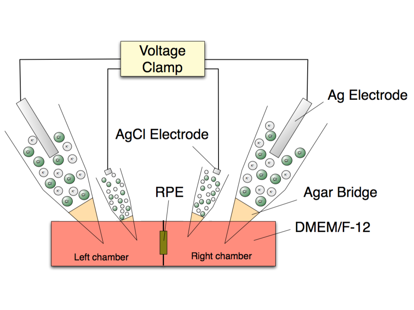
An Ussing chamber is comprised of two half chambers, which are filled with a buffer, for instance Ringer’s solution. We managed to micro-surgically isolate the retinal pigment epithelium (RPE) from the rat eye, which is ~2 mm in diameter.
The RPE is a key building block of the outer blood-retinal barrier (oBRB). The oBRB is compromised in exudative age-related macula degeneration (AMD) or in diabetic macula edema (DME). This happens for instance, when blood vessels cross the oBRB and leak or bleed into the inner eye. Other conditions, where changes in the oBRB play a role, include diabetic macula edema (DME).
We have succeeded in recording stable TEER values for over 2 hours from single explants. Beyond that time period, the cells at the edge of the explant leak current, which leads to a gradual decrease in TEER. However, within the first 60 minutes, we can reliably perform experiments in rat-eye explants.
This is a unique experimental setup that allows functional studies of genuine RPE ex vivo.